Under the microscope–.
The 2019 Olympus Global Image of the Year honorees find charm under the microscope.
Jennifer Ouellette
–
.
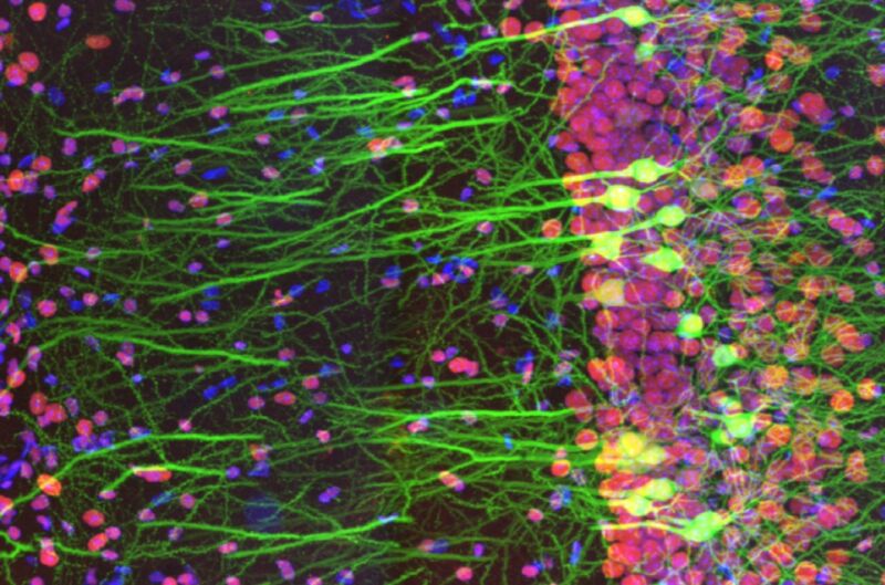
Enlarge / Information from the winning entry in the first Olympus Global Picture of the Year Life Science Light Microscopy Award. It reveals immunostaining of a mouse-brain slice with 2 fluorophores.
.
For several years now, we have actually frequently featured the winners of Nikon’s yearly Little World microscopy contest. Now, Olympus has actually gone into the artistic imaging arena with its very first Global Image of the Year Award.
While it might not matter for clinical functions, a compelling microscope image depends on things like structure, lighting, direct exposure, and more.
Spain’s Ainara Pintor snagged the top honor from over 400 submissions with her beautiful image of an immunostained mouse-brain slice, entitled Neurogarden The image concentrates on the hippocampus area of a single piece, however there are more than 70 million nerve cells in the mouse brain as a whole, according to Pintor. Howard Vindin of Australia won the regional prize for Asia-Pacific by capturing an autofluorescence picture of a mouse embryo. US entrant Tagide de Carvalho won the local award for the Americas with his colorful image of a tardigrade. The local winner for Europe, the Middle East, and Africa was the UK’s Alan Prescott, for his image catching the frozen area of a mouse’s head.
Respectable discusses consisted of striking microscopic pictures of photonic crystals in insect scales, crystallized amino acids, desert locust wings, and opal ingrained in iron sandstone, among others. Plainly, the field of photomicroscopy is still drawing in top-notch talent.
-
International Winner: immunostaining of a mouse brain slice with two fluorophores
-
Regional Winner for the Americas: it’s a vibrant tardigrade!
Tagide de Carvalho/Olympus -
Asia-Pacific Regional Winner: Autofluorescence picture of a mouse embryo.
Howard Vindin/Olympus -
EMEA Regional Winner: frozen area of a mouse’s head.
Alan Prescott/Olympus -
Honorable Mention: mouse spine.
Tong Zhang/Olympus -
Honorable Reference: picture of 3D depth color-coded reconstruction of confocal images of microtubules in monkey fibroblast cells.
Daniela Malide/Olympus -
Photonic crystals in bugs (beetles and weevils) FTW!
Rudolf Buechi/Olympus -
Preparation of amino acids crystallized out of an ethanol option.
Justin Zoll/Olympus -
The desert locust is one example of an insect types that has actually evolved collapsible wings, the much better to keep them tidy and intact. This image is called A Road in the Sky because “veins look like roadways and spines on [the] wing membrane resemble stars.”
Hamed Rajabi/Olympus -
Green gem product, prase opal, magnified through a microscopic lense, looks like an aerial shoreline view. The brown locations are iron sandstone host rock.
Nathan Renfro/Olympus -
Tiny picture of fruit fly brain.
Martin Hailstone/Olympus -
Inflorescence of developing flower buds expressing fluorescent reporters (cyan), with cell walls stained red.
Nat Prunet/Olympus -
The ovary of a gall-inducing wasp revealing the eggs.
Ming-Der Lin/Olympus